


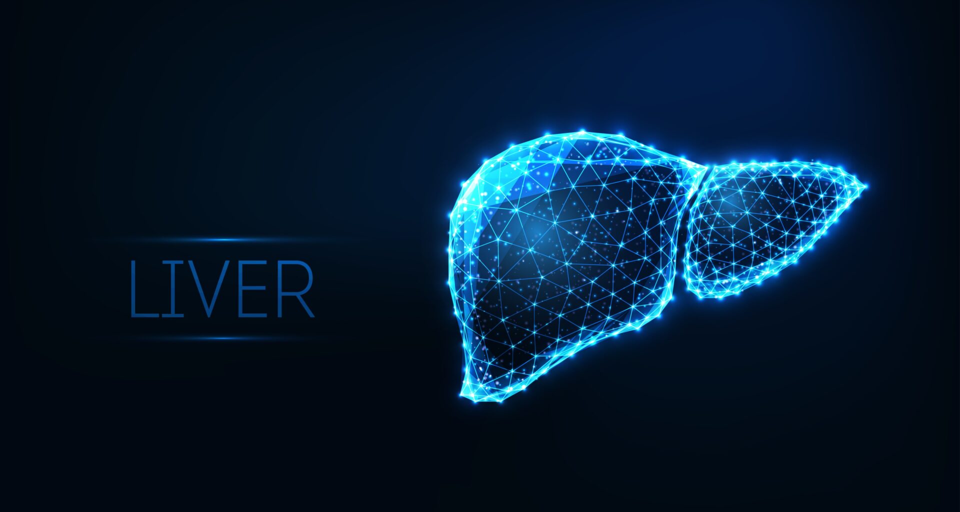

What is Liver?
The liver is the largest internal organ, and is significant for filtering and regulating the blood flow in the body. It is located in the upper right portion of the abdominal cavity, on top of the stomach. It plays a key role in the digestion of food. The liver works on the following functions:
- Removing blood from the intestines and purifying it
- Processing and preserving essential nutrients that the intestines have ingested
- Transforming nutrients into energy or materials required for tissue growth
- to make bileto help digest fat that comes from food
- to store glycogen (sugar), which the body uses for energy
- to filter harmful substances from the blood so they can be passed from the body in stools and urine

What is Liver Cancer?
Liver cancer is also known as primary liver cancer. The disease in which the tissues of the liver develop malignant (cancer) cells is called primary liver cancer. It is not the same as cancer which starts in another part of the body and travels to the liver. Instead, it is based on the location of cancer’s onset. For instance, cancers that originated in the pancreas, colon, stomach, breast, lung, or another organ and have moved to the liver are still referred to by their original organ names. This is significant to understand since primary liver cancer and metastatic disease are treated differently.
Types of Liver Cancer
Adults can develop a number of different primary liver cancers. They bear the name of the cell type from which cancer originates.
Hepatocellular Carcinoma (HCC)
Hepatocellular carcinoma can grow in a variety of ways. Some spread growths resembling tentacles throughout the liver. Some begin as a single tumour that later spreads to additional liver regions as the disease progresses. Others form nodules in various locations throughout the liver. There are times when a pattern is unclear.


Cholangiocarcinoma (Bile Duct Cancer)
Cancel cells in the liver’s bile duct lead to cholangiocarcinomas. A skinny tube called the bile duct runs from the liver to the small intestine. The bile duct begins as a series of smaller tubes that connect inside the liver.
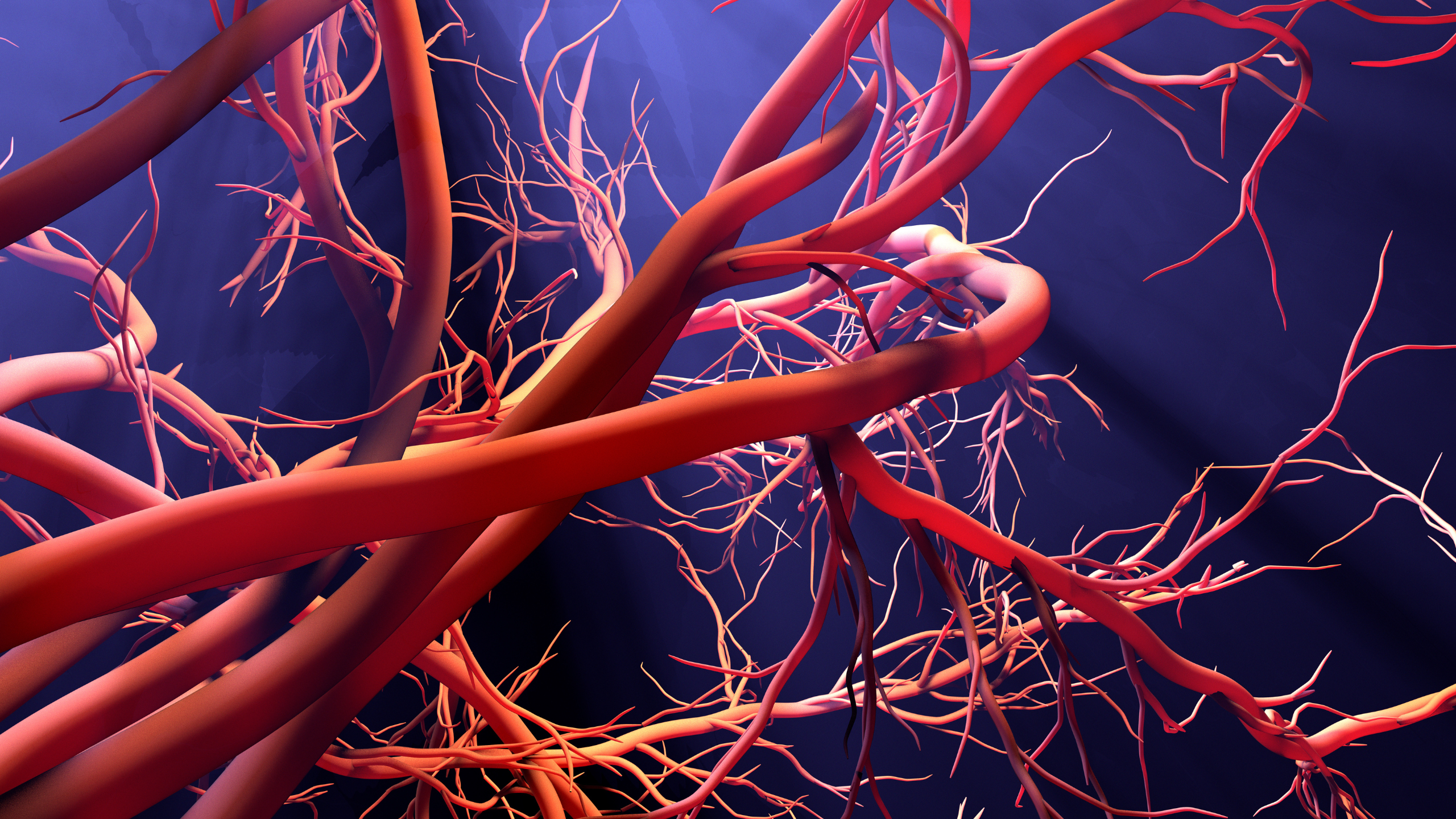
3. Angiosarcoma
Angiosarcoma develops in the blood vessels of the liver and starts spreading rapidly. It often affects the skin and may appear as a bruise-like lesion that grows over time. Angiosarcoma is a rare type of cancer that forms in the lining of the blood vessels and lymph vessels.
Causes of Liver Cancer
Liver cancer has a lot of risk factors related to it. Not everyone who possesses one or more of these risk factors will get the disease, and some individuals without any known risk factors will also become ill. The following are the risk factors for liver cancer:
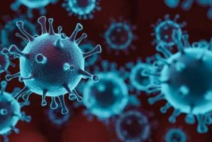
Hepatitis B virus (HBV) infection:
Blood, sperm, and other bodily fluids can all carry the HBV virus. The virus may be transferred from mother to kid after childbirth, via sexual contact, or through the sharing of drug injection needles. It may result in liver inflammation (swelling) and malignancy. The prevalence of HBV infection is declining as a result of routine infant HBV immunisation.
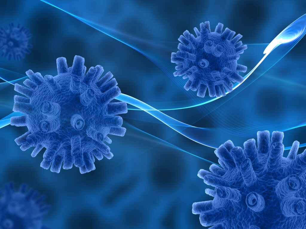
Hepatitis C virus (HCV) infection
Blood can be used to spread HCV. Sharing needles used for injecting drugs or, less frequently, sexual contact can spread the illness. It used to be transferred through organ or blood transplants as well. The danger of contracting the virus from blood transfusions has significantly decreased because blood banks now screen all donated blood for HCV. Cirrhosis brought on by it may develop into liver cancer.

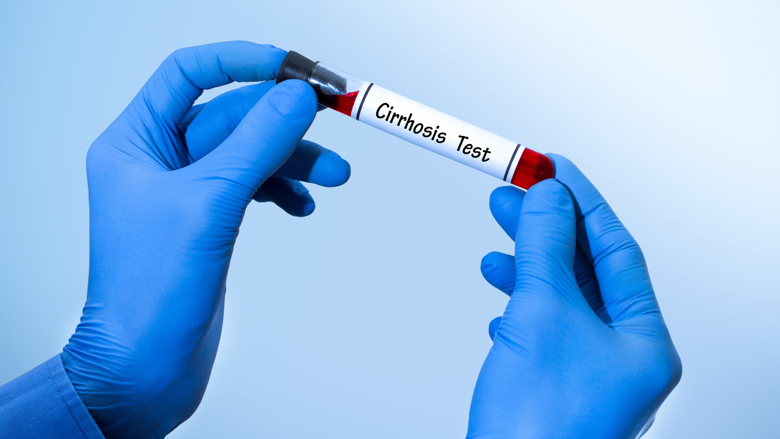
Cirrhosis
People who have cirrhosis, a condition in which healthy liver tissue is replaced by scar tissue, have a higher chance of developing liver cancer. The liver can not function properly because of the scar tissue, which prevents blood from flowing through it. The major causes of cirrhosis are alcohol consumption and chronic hepatitis infections.

Alcohol
Excessive use of alcohol can lead to cirrhosis, which increases the chance of developing liver cancer. Heavy drinkers without cirrhosis can still get liver cancer. In comparison to heavy alcohol users who do not have cirrhosis, they have a tenfold increased risk of developing liver cancer.

Smoking
Cigarette smoking has been linked to a higher risk of liver cancer. The risk increases with the number of cigarettes smoked per day and the number of years the person has smoked.
Symptoms of Liver Cancer
You must know when to visit a doctor if you notice these common symptoms of liver cancer –
- A swollen abdomen
- A rough spot directly below the ribcage on the right side
- Irritation on the right side of the upper abdomen
- Pain in the back or near the right shoulder blade
- Easy bleeding or bruising
- unexpected fatigue or weakness
- Feeling full with small portions of meals or losing appetite
Diagnoses for Liver Cancer
Physical and health examination:
An individual’s health will be evaluated through a physical examination of their body, which will also look for any abnormalities or tumours that might be symptoms of the disease. Additionally, a history of the patient’s health practises, diseases, and treatments in the past will be recorded.

Alpha-fetoprotein (AFP) tumour marker test:
Tumor markers are released into the blood by organs, tissues, or tumor cells in the body. An increased level of AFP in the blood may be a sign of liver cancer. Other cancers and certain noncancerous conditions, including cirrhosis and hepatitis, may also increase AFP levels. Sometimes the AFP level is normal even when there is liver cancer.
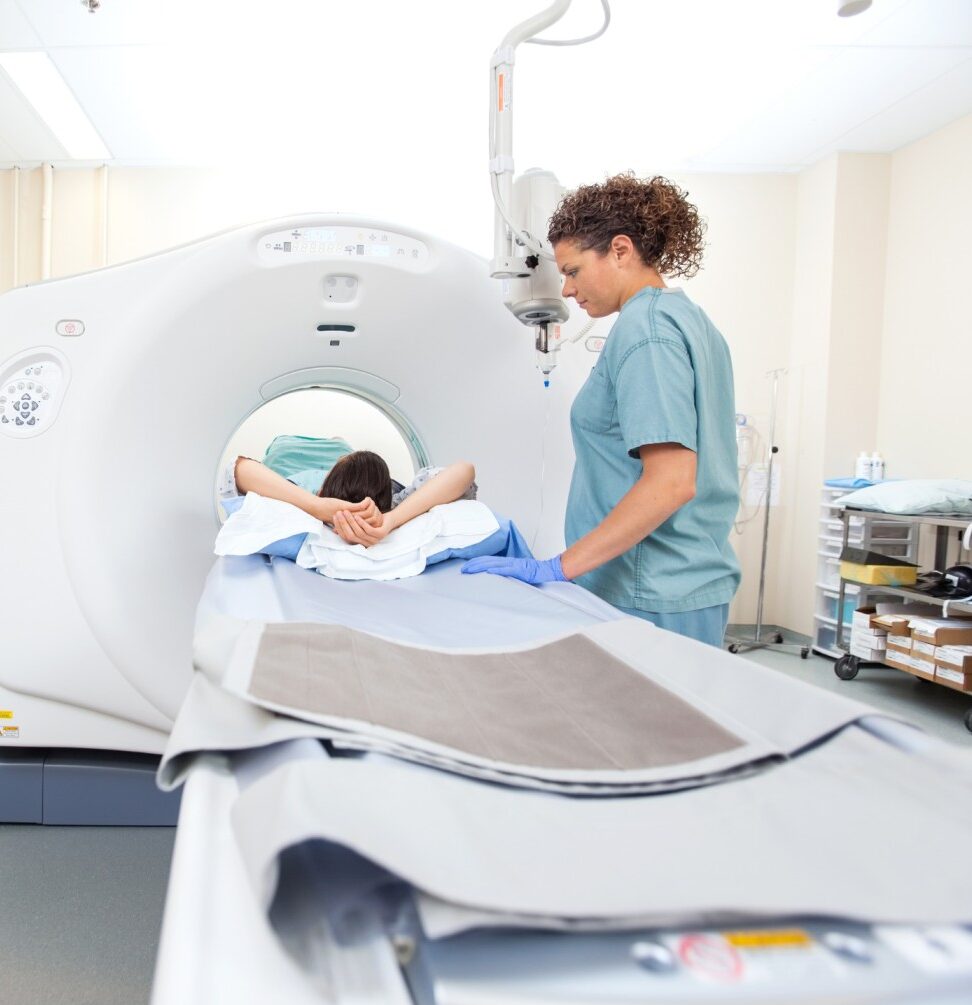
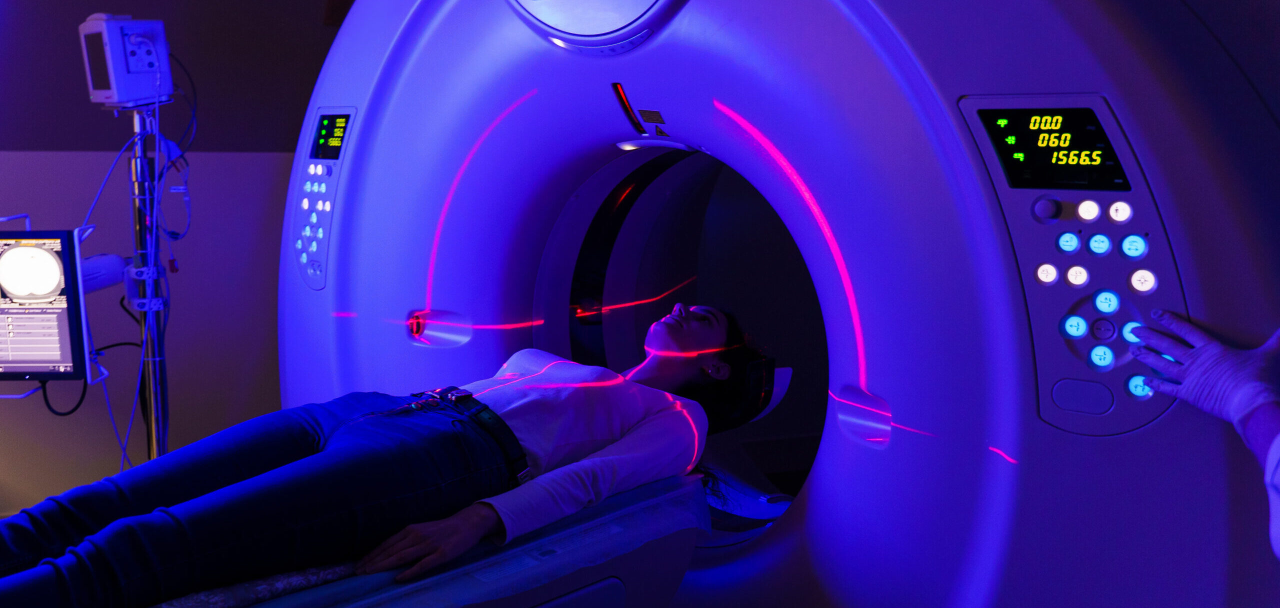
CT scan (CAT scan):
This procedure uses a computer linked to an x-ray machine to make a series of detailed pictures of areas inside the body, such as the abdomen, taken from different angles. A dye may be injected into a vein or swallowed to help the organs or tissues show up more clearly. This procedure is also called computed tomography, computerized tomography, or computerized axial tomography. Images may be taken at three different times after the dye is injected, to get the best picture of abnormal areas in the liver. This is called triple-phase CT. A spiral or helical CT scan makes a series of very detailed pictures of areas inside the body using an x-ray machine that scans the body in a spiral path.
Preventions and Treatments for Bladder Cancer
Preventions to be considered are as follows
Cancer prevention aims to lessen the likelihood of developing the disease. A group or population’s risk of developing cancer is reduced by preventing it. Hopefully, this will reduce the number of cancer-related fatalities.


Getting the hepatitis B vaccine:
Preventing HBV infection (by being vaccinated for HBV as a newborn) has been shown to lower the risk of liver cancer in children. It is not yet known if being vaccinated lowers the risk of liver cancer in adults.
Getting treatment for chronic hepatitis B infection:
Treatment options for people with chronic HBV infection include interferon and nucleos(t)ide analog therapy. These treatments may reduce the risk of developing liver cancer.
Reducing exposure to aflatoxin B1:
Replacing foods that contain high amounts of aflatoxin B1 with foods that contain a much lower level of the poison can reduce the risk of liver cancer.
Treatments required for Liver Cancer

Surgery is the removal of the tumour along with part of the surrounding healthy tissue. In particular, for patients with healthy livers and tumours that can be safely removed from a small amount of the liver, it is probably the most effective disease-directed treatment. If the tumour has moved outside of the liver, the liver is excessively damaged, or the patient is suffering from other significant conditions, surgery may not be an option.
Two types of surgeries to treat liver cancer are:
- Hepatectomy: When a portion of the liver is removed, the surgery is called a hepatectomy. A hepatectomy can be done only if the cancer is in 1 part of the liver and the liver is working well. The remaining section of the liver takes over the functions of the entire liver. The liver may grow back to its normal size within a few weeks. A hepatectomy may not be possible if the patient has advanced cirrhosis, even if the tumour is small.
- Liver transplantation: Sometimes, liver transplantation can be done. This is a surgery in which the patient’s liver is removed and replaced by healthy liver tissue from a donor. This procedure is possible only when specific criteria are met, including having a certain tumour size and number of tumors and whether a suitable donor is found. These criteria are usually either having a single tumor that is 5 cm or smaller or having 3 or fewer tumors, all of which are smaller than 3 cm. It is important to note that the number of available donor livers is very limited, so transplantation is not always an option.

Ablation therapy removes or destroys tissue. Different types of ablation therapy are used for liver cancer:
- Radiofrequency ablation: Special needles are inserted directly through the skin or through an incision in the abdomen to reach the tumour. High-energy radio waves heat the needles and tumors which kills cancer cells.
- Microwave therapy: The tumour is exposed to high temperatures created by microwaves. This can damage and kill cancer cells or make them more sensitive to the effects of radiation and certain anticancer drugs.
- Percutaneous ethanol injection: A small needle is used to inject ethanol (pure alcohol) directly into a tumor to kill cancer cells. Several treatments may be needed. Usually local anesthesia is used, but if the patient has many tumors in the liver, general anesthesia may be used.
External radiation therapy uses a machine outside the body to send high-energy x-rays or other types of radiation toward the area of the body with cancer. This kills cancer cells or keeps them from growing. Certain ways of giving external radiation therapy can help keep radiation from damaging nearby healthy tissue:
- Conformal radiation therapy: Conformal radiation therapy uses a computer to make a 3-dimensional, or 3-D, picture of the tumour and shapes the radiation beams to fit the tumour. This allows a high dose of radiation to reach the tumour and causes less damage to nearby healthy tissue.
- Stereotactic body radiation therapy: Stereotactic body radiation therapy uses special equipment to place the patient in the same position for each radiation treatment. Once a day for several days, a radiation machine aims a larger-than-usual dose of radiation directly at the tumour. By having the patient in the same position for each treatment, there is less damage to nearby healthy tissue. This procedure is also called stereotactic external-beam radiation therapy and stereotaxic radiation therapy.

Targeted therapy is drug treatment that targets the cancer’s specific genes, proteins, or the tissue environment that contributes to cancer growth and survival. This type of treatment blocks the growth and spread of cancer cells and limits damage to healthy cells.
Among the anti-angiogenesis treatments are:
- with atezolizumab: In 2020, the U.S. Food and Drug Administration (FDA) approved the combination of the anti-angiogenesis targeted therapy,, with atezolizumab, an immunotherapy drug (see “Immunotherapy” below), for people with unresectable or metastatic HCC who have not received previous cancer treatment using medications. Unlike many anti-angiogenesis therapies, which are taken as pills, is given intravenously.
- Lenvatinib: In 2018, the FDA approved an anti-angiogenesis targeted therapy called lenvatinib. This drug is approved as a first treatment for HCC that cannot be removed by surgery. It is taken as a pill that is swallowed (orally).
- Sorafenib: Sorafenib is used to treat advanced HCC that cannot be completely removed with surgery. It is taken as a pill that is swallowed (orally).

Immunotherapy uses the body’s natural defences to fight cancer by improving your immune system’s ability to attack cancer cells. One common type of immunotherapy is called an immune checkpoint inhibitor. Immune checkpoint inhibitors work by blocking the pathways that would otherwise allow cancer to hide from the immune system.
Immunotherapies for HCC include:
- Nivolumab: In 2017, the FDA approved an immunotherapy called nivolumab for the treatment of HCC. Nivolumab can be used to treat people who have already been treated with sorafenib, which is a type of targeted therapy.
- Pembrolizumab: In 2018, the FDA approved the immunotherapy pembrolizumab for the treatment of people with HCC. Like nivolumab, pembrolizumab can be used to treat people who have previously been treated with sorafenib. Pembrolizumab is an immune checkpoint inhibitor.
- Nivolumab with ipilimumab (Yervoy): In 2020, the FDA approved the use of the combination of nivolumab with another immunotherapy drug called ipilimumab (Yervoy) to treat patients with HCC who have already been treated with sorafenib. Both nivolumab and ipilimumab are a type of immunotherapy called immune checkpoint inhibitors, which means they work to block the pathways that would otherwise allow cancer to hide from the immune system. Both nivolumab and ipilimumab are immune checkpoint inhibitors.
References:
What is Liver?
The liver is the largest internal organ, and is significant for filtering and regulating the blood flow in the body. It is located in the upper right portion of the abdominal cavity, on top of the stomach. It plays a key role in the digestion of food. The liver works on the following functions:
- Removing blood from the intestines and purifying it
- Processing and preserving essential nutrients that the intestines have ingested
- Transforming nutrients into energy or materials required for tissue growth
- to make bileto help digest fat that comes from food
- to store glycogen (sugar), which the body uses for energy
- to filter harmful substances from the blood so they can be passed from the body in stools and urine
What is Liver Cancer?
Liver cancer is also known as primary liver cancer. The disease in which the tissues of the liver develop malignant (cancer) cells is called primary liver cancer. It is not the same as cancer which starts in another part of the body and travels to the liver. Instead, it is based on the location of cancer’s onset. For instance, cancers that originated in the pancreas, colon, stomach, breast, lung, or another organ and have moved to the liver are still referred to by their original organ names. This is significant to understand since primary liver cancer and metastatic disease are treated differently.
Types of Liver Cancer
Adults can develop a number of different primary liver cancers. They bear the name of the cell type from which cancer originates.
Hepatocellular Carcinoma (HCC)
Hepatocellular carcinoma can grow in a variety of ways. Some spread growths resembling tentacles throughout the liver. Some begin as a single tumour that later spreads to additional liver regions as the disease progresses. Others form nodules in various locations throughout the liver. There are times when a pattern is unclear.
Cholangiocarcinoma (Bile Duct Cancer)
Cancel cells in the liver’s bile duct lead to cholangiocarcinomas. A skinny tube called the bile duct runs from the liver to the small intestine. The bile duct begins as a series of smaller tubes that connect inside the liver.
3. Angiosarcoma
Angiosarcoma develops in the blood vessels of the liver and starts spreading rapidly. It often affects the skin and may appear as a bruise-like lesion that grows over time. Angiosarcoma is a rare type of cancer that forms in the lining of the blood vessels and lymph vessels.
Causes of Liver Cancer
Liver cancer has a lot of risk factors related to it. Not everyone who possesses one or more of these risk factors will get the disease, and some individuals without any known risk factors will also become ill. The following are the risk factors for liver cancer:

Hepatitis B virus (HBV) infection:
Blood, sperm, and other bodily fluids can all carry the HBV virus. The virus may be transferred from mother to kid after childbirth, via sexual contact, or through the sharing of drug injection needles. It may result in liver inflammation (swelling) and malignancy. The prevalence of HBV infection is declining as a result of routine infant HBV immunisation.
Hepatitis C virus (HCV) infection
Blood can be used to spread HCV. Sharing needles used for injecting drugs or, less frequently, sexual contact can spread the illness. It used to be transferred through organ or blood transplants as well. The danger of contracting the virus from blood transfusions has significantly decreased because blood banks now screen all donated blood for HCV. Cirrhosis brought on by it may develop into liver cancer.

Cirrhosis
People who have cirrhosis, a condition in which healthy liver tissue is replaced by scar tissue, have a higher chance of developing liver cancer. The liver can not function properly because of the scar tissue, which prevents blood from flowing through it. The major causes of cirrhosis are alcohol consumption and chronic hepatitis infections.
Alcohol
Excessive use of alcohol can lead to cirrhosis, which increases the chance of developing liver cancer. Heavy drinkers without cirrhosis can still get liver cancer. In comparison to heavy alcohol users who do not have cirrhosis, they have a tenfold increased risk of developing liver cancer.
Smoking
Cigarette smoking has been linked to a higher risk of liver cancer. The risk increases with the number of cigarettes smoked per day and the number of years the person has smoked.
Symptoms of Liver Cancer
You must know when to visit a doctor if you notice these common symptoms of liver cancer –
- A swollen abdomen
- A rough spot directly below the ribcage on the right side
- Irritation on the right side of the upper abdomen
- Pain in the back or near the right shoulder blade
- Easy bleeding or bruising
- unexpected fatigue or weakness
- Feeling full with small portions of meals or losing appetite
Diagnoses for Liver Cancer
Physical and health examination:
An individual’s health will be evaluated through a physical examination of their body, which will also look for any abnormalities or tumours that might be symptoms of the disease. Additionally, a history of the patient’s health practises, diseases, and treatments in the past will be recorded.
Alpha-fetoprotein (AFP) tumour marker test:
Tumor markers are released into the blood by organs, tissues, or tumor cells in the body. An increased level of AFP in the blood may be a sign of liver cancer. Other cancers and certain noncancerous conditions, including cirrhosis and hepatitis, may also increase AFP levels. Sometimes the AFP level is normal even when there is liver cancer.

CT scan (CAT scan):
This procedure uses a computer linked to an x-ray machine to make a series of detailed pictures of areas inside the body, such as the abdomen, taken from different angles. A dye may be injected into a vein or swallowed to help the organs or tissues show up more clearly. This procedure is also called computed tomography, computerized tomography, or computerized axial tomography. Images may be taken at three different times after the dye is injected, to get the best picture of abnormal areas in the liver. This is called triple-phase CT. A spiral or helical CT scan makes a series of very detailed pictures of areas inside the body using an x-ray machine that scans the body in a spiral path.
Preventions and Treatments for Bladder Cancer
Preventions to be considered are as follows
Cancer prevention aims to lessen the likelihood of developing the disease. A group or population’s risk of developing cancer is reduced by preventing it. Hopefully, this will reduce the number of cancer-related fatalities.
Getting the hepatitis B vaccine:
Preventing HBV infection (by being vaccinated for HBV as a newborn) has been shown to lower the risk of liver cancer in children. It is not yet known if being vaccinated lowers the risk of liver cancer in adults.
Getting treatment for chronic hepatitis B infection:
Treatment options for people with chronic HBV infection include interferon and nucleos(t)ide analog therapy. These treatments may reduce the risk of developing liver cancer.
Reducing exposure to aflatoxin B1:
Replacing foods that contain high amounts of aflatoxin B1 with foods that contain a much lower level of the poison can reduce the risk of liver cancer.
Treatments required for Liver Cancer
Surgery is the removal of the tumour along with part of the surrounding healthy tissue. In particular, for patients with healthy livers and tumours that can be safely removed from a small amount of the liver, it is probably the most effective disease-directed treatment. If the tumour has moved outside of the liver, the liver is excessively damaged, or the patient is suffering from other significant conditions, surgery may not be an option.
Two types of surgeries to treat liver cancer are:
- Hepatectomy: When a portion of the liver is removed, the surgery is called a hepatectomy. A hepatectomy can be done only if the cancer is in 1 part of the liver and the liver is working well. The remaining section of the liver takes over the functions of the entire liver. The liver may grow back to its normal size within a few weeks. A hepatectomy may not be possible if the patient has advanced cirrhosis, even if the tumour is small.
- Liver transplantation: Sometimes, liver transplantation can be done. This is a surgery in which the patient’s liver is removed and replaced by healthy liver tissue from a donor. This procedure is possible only when specific criteria are met, including having a certain tumour size and number of tumors and whether a suitable donor is found. These criteria are usually either having a single tumor that is 5 cm or smaller or having 3 or fewer tumors, all of which are smaller than 3 cm. It is important to note that the number of available donor livers is very limited, so transplantation is not always an option.
Ablation therapy removes or destroys tissue. Different types of ablation therapy are used for liver cancer:
- Radiofrequency ablation: Special needles are inserted directly through the skin or through an incision in the abdomen to reach the tumour. High-energy radio waves heat the needles and tumors which kills cancer cells.
- Microwave therapy: The tumour is exposed to high temperatures created by microwaves. This can damage and kill cancer cells or make them more sensitive to the effects of radiation and certain anticancer drugs.
- Percutaneous ethanol injection: A small needle is used to inject ethanol (pure alcohol) directly into a tumor to kill cancer cells. Several treatments may be needed. Usually local anesthesia is used, but if the patient has many tumors in the liver, general anesthesia may be used.
External radiation therapy uses a machine outside the body to send high-energy x-rays or other types of radiation toward the area of the body with cancer. This kills cancer cells or keeps them from growing. Certain ways of giving external radiation therapy can help keep radiation from damaging nearby healthy tissue:
- Conformal radiation therapy: Conformal radiation therapy uses a computer to make a 3-dimensional, or 3-D, picture of the tumour and shapes the radiation beams to fit the tumour. This allows a high dose of radiation to reach the tumour and causes less damage to nearby healthy tissue.
- Stereotactic body radiation therapy: Stereotactic body radiation therapy uses special equipment to place the patient in the same position for each radiation treatment. Once a day for several days, a radiation machine aims a larger-than-usual dose of radiation directly at the tumour. By having the patient in the same position for each treatment, there is less damage to nearby healthy tissue. This procedure is also called stereotactic external-beam radiation therapy and stereotaxic radiation therapy.
Targeted therapy is drug treatment that targets the cancer’s specific genes, proteins, or the tissue environment that contributes to cancer growth and survival. This type of treatment blocks the growth and spread of cancer cells and limits damage to healthy cells.
Among the anti-angiogenesis treatments are:
- Bevacizumab with atezolizumab: In 2020, the U.S. Food and Drug Administration (FDA) approved the combination of the anti-angiogenesis targeted therapy, bevacizumab, with atezolizumab, an immunotherapy drug (see “Immunotherapy” below), for people with unresectable or metastatic HCC who have not received previous cancer treatment using medications. Unlike many anti-angiogenesis therapies, which are taken as pills, bevacizumab is given intravenously.
- Lenvatinib: In 2018, the FDA approved an anti-angiogenesis targeted therapy called lenvatinib. This drug is approved as a first treatment for HCC that cannot be removed by surgery. It is taken as a pill that is swallowed (orally).
- Sorafenib: Sorafenib is used to treat advanced HCC that cannot be completely removed with surgery. It is taken as a pill that is swallowed (orally).
Immunotherapy uses the body’s natural defences to fight cancer by improving your immune system’s ability to attack cancer cells. One common type of immunotherapy is called an immune checkpoint inhibitor. Immune checkpoint inhibitors work by blocking the pathways that would otherwise allow cancer to hide from the immune system.
Immunotherapies for HCC include:
- Nivolumab: In 2017, the FDA approved an immunotherapy called nivolumab for the treatment of HCC. Nivolumab can be used to treat people who have already been treated with sorafenib, which is a type of targeted therapy.
- Pembrolizumab: In 2018, the FDA approved the immunotherapy pembrolizumab for the treatment of people with HCC. Like nivolumab, pembrolizumab can be used to treat people who have previously been treated with sorafenib. Pembrolizumab is an immune checkpoint inhibitor.
- Nivolumab with ipilimumab (Yervoy): In 2020, the FDA approved the use of the combination of nivolumab with another immunotherapy drug called ipilimumab (Yervoy) to treat patients with HCC who have already been treated with sorafenib. Both nivolumab and ipilimumab are a type of immunotherapy called immune checkpoint inhibitors, which means they work to block the pathways that would otherwise allow cancer to hide from the immune system. Both nivolumab and ipilimumab are immune checkpoint inhibitors.
References: