


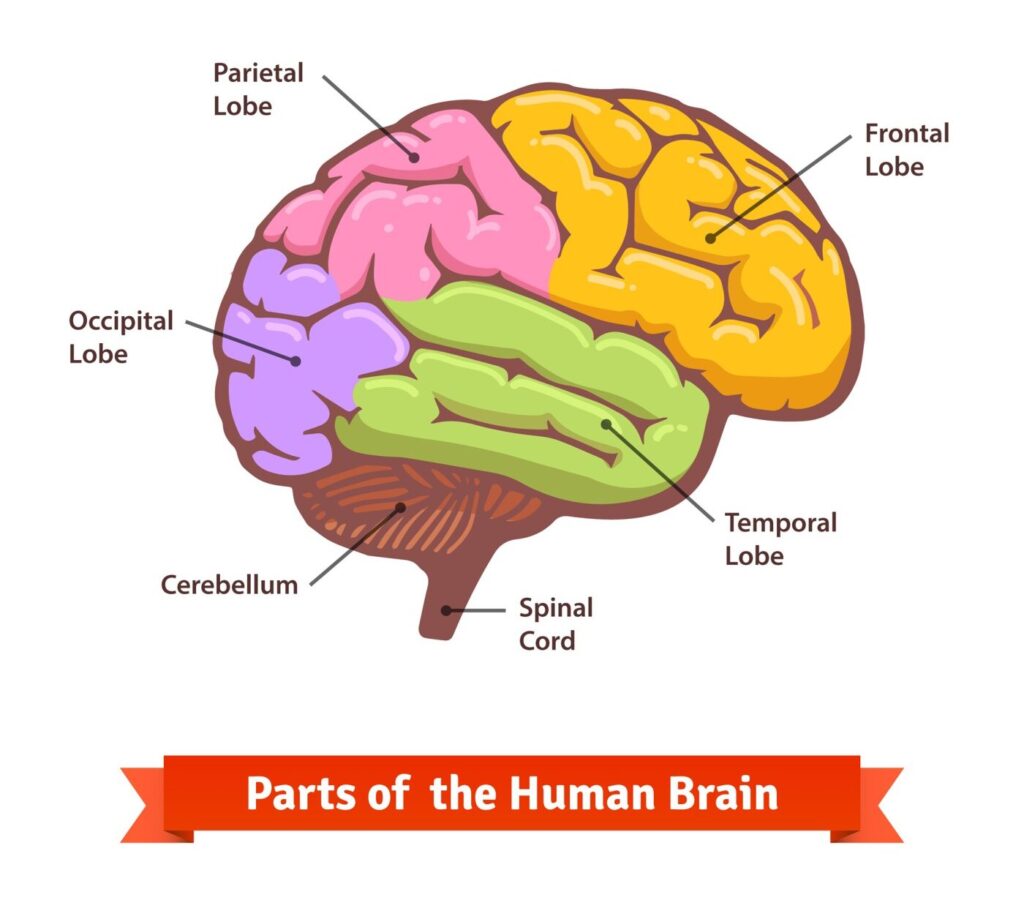
What is the Brain?
The brain is an incredibly complex and important organ in the human body. It is the control center of the nervous system and plays a critical role in nearly every aspect of human life, from regulating bodily functions like breathing and heartbeat to processing thoughts and emotions.
The brain is composed of billions of specialized cells called neurons, which are responsible for transmitting electrical and chemical signals throughout the brain and the rest of the body. These signals are essential for a wide range of functions, including sensory perception, movement, learning, memory, and emotions.

What is Brain Tumour?
A brain tumour is a mass or growth of abnormal cells in the brain that can interfere with normal brain function. Brain tumours can be either cancerous (malignant) or non-cancerous (benign), and they can either originate in the brain (primary brain tumours) or spread to the brain from other parts of the body (metastatic brain tumours).
Primary Brain Tumour:
A primary brain tumour is a tumour that starts in the brain. A primary brain tumour is often described as “low grade” or “high grade.” A low-grade tumour generally grows slowly, but it can turn into a high-grade tumour. A high-grade tumour is more likely to grow faster.
In adults, secondary brain tumours, also called brain metastases, are much more common than primary tumours.
Secondary Brain Tumour:
A secondary brain tumour is a cancerous tumour that started in another part of the body, such as the breast, lung, or colon, and then spread to the brain. A secondary brain tumour may also be called metastatic cancer or brain metastasis.
If cancer spreads to the meninges and the cerebrospinal fluid (CSF), it is called leptomeningeal metastases or neoplastic meningitis. This condition occurs more commonly in people with leukemia, lymphoma, melanoma, breast cancer, or lung cancer.

Types of Primary Brain Tumour
There are many types of primary brain tumours. Some cannot be assigned an exact type because the tumour’s location makes it too difficult to remove for full testing.
Gliomas
As a group, gliomas are one of the most common types of brain tumours. While the exact origin of gliomas is still unknown, they are thought to grow from glial cells or glial precursor cells. A glial cell is a type of supportive cell in the brain. The main types of supportive cells in the brain include astrocytes, oligodendrocytes, and ependymal cells. Gliomas may be considered astrocytoma, oligodendroglioma, or ependymoma.
Currently, the types of gliomas include:
- Astrocytoma: Astrocytoma is the most common type of glioma. Astrocytoma cells look like glial cells called astrocytes that are found in the cerebrum or cerebellum. Astrocytoma in children is more common than astrocytoma in adults.
- Oligodendroglioma: Oligodendroglioma is a tumour whose cells look like glial cells called oligodendrocytes. These cells are responsible for making myelin. Myelin surrounds the nerves and is rich in protein and fatty substances called lipids.
- Ependymoma: Ependymoma commonly begins in the passageways in the brain where CSF is made and stored. In adults, they occur more often in the spine and can also be of the myxopapillary subtype.
- Brain stem glioma: A brain stem glioma begins in the glial cells in the brain stem.
Non-glioma tumours
Non-glioma tumours are tumours that arise from cells in the brain that are not glial cells. Types of non-glioma tumours include:
- Meningioma: Meningioma is the most common primary brain tumour. It begins in the meninges and is most often noncancerous. Meningioma can cause serious symptoms if it grows and presses on the brain or spinal cord or grows into the brain tissue.
- Pineal gland and pituitary gland tumours: These are tumours that start in the pineal gland and pituitary gland.
- Primary CNS lymphoma: This is a form of lymphoma. Lymphoma is cancer that begins in the lymphatic system. Primary CNS lymphoma starts in the brain and can spread to the spinal fluid and eyes.
- Medulloblastoma: Medulloblastoma is thought to start from a specific type of cell in the cerebellum. These cells are called cerebellar granule progenitor cells. It is most common in children and is usually cancerous, often spreading throughout the CNS.
- Craniopharyngioma: Craniopharyngioma is a benign tumour that begins near the pituitary gland located near the base of the brain. These tumours are rare.
- Schwannoma: Schwannoma is a rare tumour that begins in the nerve sheath, or the lining of the nerves. It may often occur in the vestibular nerve, which is a nerve in the inner ear that helps control balance. It is typically noncancerous.
Risk Factors of Brain Tumour
The exact causes of brain tumours are not fully understood, but several risk factors have been identified that may increase the likelihood of developing a brain tumour. These risk factors include –
Exposure to radiation
Family history may signify either a genetic abnormality or shared environmental factors, or a combination of these factors. About 15% of all colorectal cancers occur in patients with a history of colorectal cancer in first-degree relatives.

Genetic disorders
Lack of physical activity and a sedentary lifestyle can increase the risk of colorectal cancer.

Family history
Being overweight or obese increases the risk of colorectal cancer, especially in men.

Age
Men and women smoking during the previous 20 years have three times the relative risk for small adenomas (<1 cm) but not for larger ones. Smoking for >20 years was associated with 2.5 times the relative risk for larger adenomas.

Immune system disorders
Certain conditions that affect the immune system, such as HIV/AIDS, may increase the risk of developing a brain tumour.

Chemical exposure
Exposure to certain chemicals, such as pesticides or industrial solvents, may increase the risk of developing a brain tumour.
Symptoms of Brain Tumour
In its early stages, colorectal cancer may not produce any noticeable symptoms. However, as cancer grows and spreads, it can cause several symptoms. Here are some of the common symptoms of colorectal cancer –
- Changes in bowel habits, such as diarrhoea or constipation last for more than a few days.
- Blood in the stool or rectal bleeding, may appear as bright red or very dark.
- Abdominal pain or discomfort, including cramping or bloating.
- Unexplained weight loss.
- Fatigue or weakness.
- Narrow stools.
- Feeling like you need to have a bowel movement that is not relieved by doing so.
- Nausea or vomiting.
These symptoms can be caused by other conditions, such as haemorrhoids or inflammatory bowel disease. However, if you experience any of these symptoms, especially if they persist for more than a few days, you should consult your healthcare provider for an evaluation. Early detection of colorectal cancer is key to successful treatment and improved outcomes. Screening tests such as colonoscopy can help detect cancer at an early stage, even before symptoms appear.
Diagnoses of Brain Tumour
The diagnosis of colorectal cancer typically involves several steps, including –
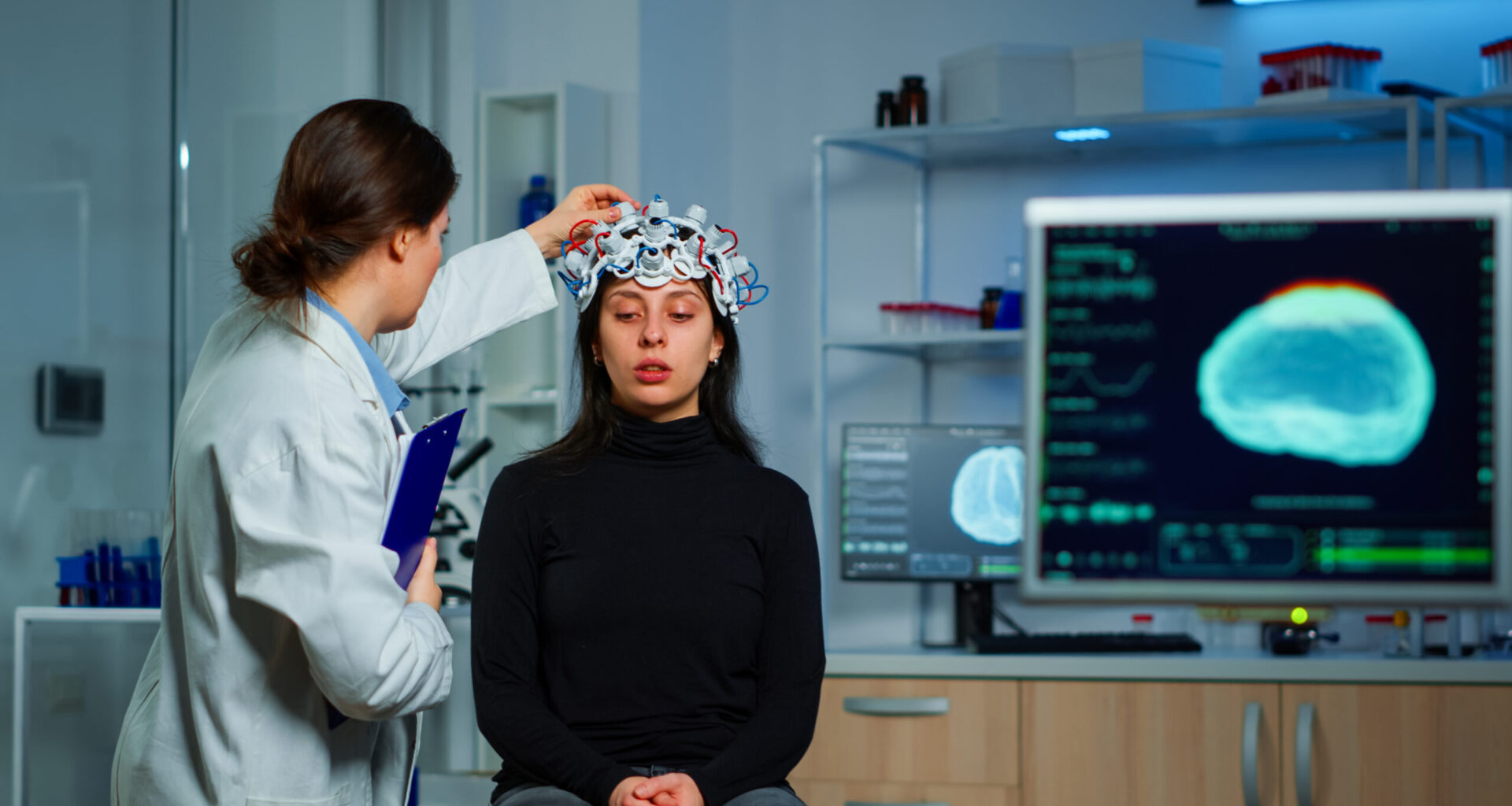
Magnetic resonance imaging (MRI):
An MRI uses magnetic fields, not x-rays, to produce detailed images of the body. MRI can be used to measure the tumour’s size. A special dye called a contrast medium is given before the scan to create a clearer picture. This dye can be injected into a patient’s vein or given as a pill or liquid to swallow. MRIs create more detailed pictures than CT scans and are the preferred way to diagnose a brain tumour. The MRI may be of the brain, spinal cord, or both, depending on the type of tumour suspected and the likelihood that it will spread in the CNS.

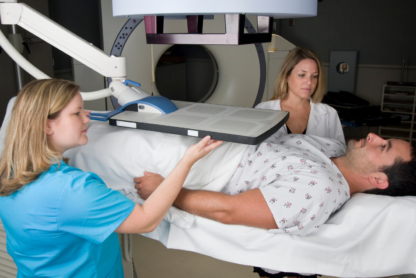
CT scan:
A CT scan takes pictures of the inside of the body using x-rays taken from different angles. A computer combines these pictures into a detailed, 3-dimensional image that shows any abnormalities or tumours. A CT scan can help find bleeding and enlargement of the fluid-filled spaces in the brain, called ventricles. Changes to bone in the skull can also be seen on a CT scan, and it can be used to measure a tumour’s size. A CT scan may also be used if the patient cannot have an MRI, such as if the person has a pacemaker for their heart.
Tissue sampling/biopsy:
A sample of the tumour’s tissue is usually needed to make a final diagnosis. A biopsy is the removal of a small amount of tissue for examination under a microscope and is the only definitive way a brain tumour can be diagnosed. A pathologist then analyzes the sample(s). A pathologist is a doctor who specializes in interpreting laboratory tests and evaluating cells, tissues, and organs to diagnose disease. A biopsy can be done as part of surgery to remove the entire tumour.

Positron emission tomography (PET) or PET-CT scan:
A PET scan is used at first to find out more about a tumour while a patient is receiving treatment. It may also be used if the tumour comes back after treatment. A PET scan is usually combined with a CT scan (see above), called a PET-CT scan. However, you may hear your doctor refer to this procedure just as a PET scan. A PET scan is a way to create pictures of organs and tissues inside the body using various substances, such as sugars or proteins. A small amount of a radioactive substance is injected into the patient’s body. This substance is taken up by cells that are actively dividing. Because tumour cells are more likely to be actively dividing, they absorb more of the radioactive substance.
Cerebral arteriogram (cerebral angiogram):
A cerebral arteriogram is an x-ray, or series of x-rays, of the head that shows the arteries in the brain. X-rays are taken after a special dye called a contrast medium is injected into the main arteries of the patient’s head.

Preventions and Treatments for Brain Tumour
Preventions to be considered are as follows
There are several measures that can help reduce the risk of developing brain tumour –


Limiting exposure to radiation: Exposure to high levels of ionizing radiation, such as radiation used in cancer treatment or from atomic bombs, has been linked to an increased risk of brain tumours. Limiting your exposure to radiation as much as possible can help to reduce your risk.
- Protecting your head from injury: Traumatic brain injury has been linked to an increased risk of developing certain types of brain tumours. Wearing a helmet when participating in high-risk activities such as sports, and taking steps to prevent falls, can help to reduce your risk of head injury.
- Reducing exposure to certain chemicals: Exposure to certain chemicals, such as pesticides or industrial solvents, has been linked to an increased risk of brain tumours. Taking steps to reduce your exposure to these chemicals can help to reduce your risk.
Treatments required for Brain Tumour
The treatment for a brain tumour depends on several factors, including the type, location, size, and grade of the tumour, as well as the patient’s age and overall health. Treatment options may include –

Surgery is the removal of the tumour and some surrounding healthy tissue during an operation. It is usually the first treatment used for a brain tumour. It is often the only treatment needed for a low-grade brain tumour. Removing the tumour can improve neurological symptoms, provide tissue for diagnosis and genetic analysis, help make other brain tumour treatments more effective, and, in many instances, improve the prognosis of a person with a brain tumour.
A neurosurgeon is a doctor who specializes in surgery on the brain and spinal column. Surgery to the brain requires the removal of part of the skull, a procedure called a craniotomy. After the surgeon removes the tumour, the patient’s own bone will be used to cover the opening in the skull.
There have been rapid advances in surgery for brain tumours, including the use of cortical mapping, enhanced imaging, and fluorescent dyes.
- Cortical mapping allows doctors to identify areas of the brain that control the senses, language, and motor skills.
- Enhanced imaging devices give surgeons more tools to plan and perform surgery. For example, computer-based techniques, such as image guided surgery (IGS), help surgeons map out the location of the tumour very accurately. However, this is a very specialized technique that may not be widely available.
- A fluorescent dye, called 5-aminolevulinic acid, can be given by mouth the morning before surgery. This dye is taken up by tumour cells. Doctors can use a special microscope and light to see the cells that have taken up the dye during the surgery. This helps doctors safely remove as much of the tumour as possible.

Radiation therapy is the use of high-energy x-rays or other particles to destroy tumour cells. Doctors may use radiation therapy to slow or stop the growth of a brain tumour. It is typically given after surgery and possibly along with chemotherapy. A doctor who specializes in giving radiation therapy to treat a tumour is called a radiation oncologist. The most common type of radiation treatment is called external-beam radiation therapy, which is radiation given from a machine outside the body. When radiation treatment is given using implants, it is called internal radiation therapy or brachytherapy. A radiation therapy regimen, or schedule, usually consists of a specific number of treatments given over a set period of time.
External-beam radiation therapy can be directed at a brain tumour in the following ways:
- Conventional radiation therapy. The treatment location is determined based on anatomic landmarks and x-rays. In certain situations, such as whole-brain radiation therapy for brain metastases, this technique is appropriate. For more precise targeting, different techniques are needed. The amount of radiation given depends on the tumour’s grade.
- 3-dimensional conformal radiation therapy (3D-CRT). Using images from CT and MRI scans (see Diagnosis), a 3-dimensional model of the tumour and healthy tissue surrounding the tumour is created on a computer. This model can be used to aim the radiation beams directly at the tumour, sparing the healthy tissue from high doses of radiation therapy.
- Intensity modulated radiation therapy (IMRT). IMRT is a type of 3D-CRT (see above) that can more directly target a tumour. It can deliver higher doses of radiation to the tumour while giving less to the surrounding healthy tissue. In IMRT, the radiation beams are broken up into smaller beams and the intensity of each of these smaller beams can be changed. This means that the more intense beams, or the beams giving more radiation, can be directed only at the tumour.
- Proton therapy. Proton therapy is a type of external-beam radiation therapy that uses protons rather than x-rays. At high energy, protons can destroy tumour cells. Proton beam therapy is typically used for tumours when less radiation is needed because of the location. This includes tumours that have grown into nearby bone, such as the base of skull, and those near the optic nerve.
- Stereotactic radiosurgery. Stereotactic radiosurgery is the use of a single, high dose of radiation given directly to the tumour and not healthy tissue. It works best for a tumour that is only in 1 area of the brain and certain noncancerous tumours. It can also be used when a person has more than 1 metastatic brain tumour.
- Fractionated stereotactic radiation therapy. Radiation therapy is delivered with stereotactic precision but divided into small daily doses called fractions and given over several days or weeks, in contrast to the 1-day radiosurgery. This technique is used for tumours located close to sensitive structures, such as the optic nerves or brain stem.

Chemotherapy is the use of drugs to destroy tumour cells, usually by keeping the tumour cells from growing, dividing, and making more cells.
A chemotherapy regimen, or schedule, usually consists of a specific number of cycles given over a set period of time. A patient may receive 1 drug at a time or a combination of different drugs given at the same time. The goal of chemotherapy can be to destroy tumour cells remaining after surgery, slow a tumour’s growth, or reduce symptoms.
As explained above, chemotherapy to treat a brain tumour is typically given after surgery and possibly with or after radiation therapy, particularly if the tumour has come back after initial treatment.
Some drugs are better at going through the blood-brain barrier. These are the drugs often used for a brain tumour.
- Gliadel wafers are a way to give the drug carmustine (BiCNU). These wafers are placed in the area where the tumour was removed during surgery.
- For people with glioblastoma and high-grade glioma, the latest standard of care is radiation therapy with daily low-dose temozolomide (Temodar). This is followed by monthly doses of temozolomide after radiation therapy for 6 months to 1 year.
- A combination of 3 drugs, lomustine (Gleostine), procarbazine (Matulane), and vincristine (Vincasar), has been used along with radiation therapy.

In addition to standard chemotherapy, targeted therapy is another way doctor’s use medication to treat cancer. Targeted therapy is a treatment that targets the tumour’s specific genes, proteins, or the tissue environment that contributes to a tumour’s growth and survival. This type of treatment blocks the growth and spread of tumour cells and limits the damage to healthy cells.
Not all tumours have the same targets, and some tumours may have more than 1 target. To find the most effective treatment, your doctor may run tests to identify the genes, proteins, and other factors in your tumour. This helps doctors better match each patient with the most effective treatment whenever possible. In addition, research studies continue to find out more about specific molecular targets and new treatments directed at them.
For a brain tumour, there are 2 types of targeted therapy that may be used:
- (Avastin, Mvasi) is an anti-angiogenesis therapy used to treat glioblastoma multiforme when previous treatment has not worked. Anti-angiogenesis therapy is focused on stopping angiogenesis, which is the process of making new blood vessels.
- Larotrectinib (Vitrakvi) and entrectinib (Rozlytrek) are a type of targeted therapy that is not specific to a certain type of tumour tumour but focuses on a specific genetic change called an NTRK fusion. This type of genetic change is found in a range of tumours, including some brain tumours.
References:
- Manual of Clinical Oncology by Bartosz Chmeilowski and Mary Territo
- cancer.net
What is the Brain?
The brain is an incredibly complex and important organ in the human body. It is the control center of the nervous system and plays a critical role in nearly every aspect of human life, from regulating bodily functions like breathing and heartbeat to processing thoughts and emotions.
The brain is composed of billions of specialized cells called neurons, which are responsible for transmitting electrical and chemical signals throughout the brain and the rest of the body. These signals are essential for a wide range of functions, including sensory perception, movement, learning, memory, and emotions.
What is Brain Tumour?
A brain tumour is a mass or growth of abnormal cells in the brain that can interfere with normal brain function. Brain tumours can be either cancerous (malignant) or non-cancerous (benign), and they can either originate in the brain (primary brain tumours) or spread to the brain from other parts of the body (metastatic brain tumours).
Primary Brain Tumour:
A primary brain tumour is a tumour that starts in the brain. A primary brain tumour is often described as “low grade” or “high grade.” A low-grade tumour generally grows slowly, but it can turn into a high-grade tumour. A high-grade tumour is more likely to grow faster.
In adults, secondary brain tumours, also called brain metastases, are much more common than primary tumours.
Secondary Brain Tumour:
A secondary brain tumour is a cancerous tumour that started in another part of the body, such as the breast, lung, or colon, and then spread to the brain. A secondary brain tumour may also be called metastatic cancer or brain metastasis.
If cancer spreads to the meninges and the cerebrospinal fluid (CSF), it is called leptomeningeal metastases or neoplastic meningitis. This condition occurs more commonly in people with leukemia, lymphoma, melanoma, breast cancer, or lung cancer.
Types of Primary Brain Tumour
There are many types of primary brain tumours. Some cannot be assigned an exact type because the tumour’s location makes it too difficult to remove for full testing.
Gliomas
As a group, gliomas are one of the most common types of brain tumours. While the exact origin of gliomas is still unknown, they are thought to grow from glial cells or glial precursor cells. A glial cell is a type of supportive cell in the brain. The main types of supportive cells in the brain include astrocytes, oligodendrocytes, and ependymal cells. Gliomas may be considered astrocytoma, oligodendroglioma, or ependymoma.
Currently, the types of gliomas include:
- Astrocytoma: Astrocytoma is the most common type of glioma. Astrocytoma cells look like glial cells called astrocytes that are found in the cerebrum or cerebellum. Astrocytoma in children is more common than astrocytoma in adults.
- Oligodendroglioma: Oligodendroglioma is a tumour whose cells look like glial cells called oligodendrocytes. These cells are responsible for making myelin. Myelin surrounds the nerves and is rich in protein and fatty substances called lipids.
- Ependymoma: Ependymoma commonly begins in the passageways in the brain where CSF is made and stored. In adults, they occur more often in the spine and can also be of the myxopapillary subtype.
- Brain stem glioma: A brain stem glioma begins in the glial cells in the brain stem.
Non-glioma tumours
Non-glioma tumours are tumours that arise from cells in the brain that are not glial cells. Types of non-glioma tumours include:
- Meningioma: Meningioma is the most common primary brain tumour. It begins in the meninges and is most often noncancerous. Meningioma can cause serious symptoms if it grows and presses on the brain or spinal cord or grows into the brain tissue.
- Pineal gland and pituitary gland tumours: These are tumours that start in the pineal gland and pituitary gland.
- Primary CNS lymphoma: This is a form of lymphoma. Lymphoma is cancer that begins in the lymphatic system. Primary CNS lymphoma starts in the brain and can spread to the spinal fluid and eyes.
- Medulloblastoma: Medulloblastoma is thought to start from a specific type of cell in the cerebellum. These cells are called cerebellar granule progenitor cells. It is most common in children and is usually cancerous, often spreading throughout the CNS.
- Craniopharyngioma: Craniopharyngioma is a benign tumour that begins near the pituitary gland located near the base of the brain. These tumours are rare.
- Schwannoma: Schwannoma is a rare tumour that begins in the nerve sheath, or the lining of the nerves. It may often occur in the vestibular nerve, which is a nerve in the inner ear that helps control balance. It is typically noncancerous.
Risk Factors of Brain Tumour
The exact causes of brain tumours are not fully understood, but several risk factors have been identified that may increase the likelihood of developing a brain tumour. These risk factors include –
Exposure to radiation
Family history may signify either a genetic abnormality or shared environmental factors, or a combination of these factors. About 15% of all colorectal cancers occur in patients with a history of colorectal cancer in first-degree relatives.
Genetic disorders
Lack of physical activity and a sedentary lifestyle can increase the risk of colorectal cancer.
Family history
Being overweight or obese increases the risk of colorectal cancer, especially in men.
Age
Men and women smoking during the previous 20 years have three times the relative risk for small adenomas (<1 cm) but not for larger ones. Smoking for >20 years was associated with 2.5 times the relative risk for larger adenomas.
Immune system disorders
Certain conditions that affect the immune system, such as HIV/AIDS, may increase the risk of developing a brain tumour.
Chemical exposure
Exposure to certain chemicals, such as pesticides or industrial solvents, may increase the risk of developing a brain tumour.
Symptoms of Brain Tumour
In its early stages, colorectal cancer may not produce any noticeable symptoms. However, as cancer grows and spreads, it can cause several symptoms. Here are some of the common symptoms of colorectal cancer –
- Changes in bowel habits, such as diarrhoea or constipation last for more than a few days.
- Blood in the stool or rectal bleeding, may appear as bright red or very dark.
- Abdominal pain or discomfort, including cramping or bloating.
- Unexplained weight loss.
- Fatigue or weakness.
- Narrow stools.
- Feeling like you need to have a bowel movement that is not relieved by doing so.
- Nausea or vomiting.
These symptoms can be caused by other conditions, such as haemorrhoids or inflammatory bowel disease. However, if you experience any of these symptoms, especially if they persist for more than a few days, you should consult your healthcare provider for an evaluation. Early detection of colorectal cancer is key to successful treatment and improved outcomes. Screening tests such as colonoscopy can help detect cancer at an early stage, even before symptoms appear.
Diagnoses of Brain Tumour
The diagnosis of colorectal cancer typically involves several steps, including –
Magnetic resonance imaging (MRI):
An MRI uses magnetic fields, not x-rays, to produce detailed images of the body. MRI can be used to measure the tumour’s size. A special dye called a contrast medium is given before the scan to create a clearer picture. This dye can be injected into a patient’s vein or given as a pill or liquid to swallow. MRIs create more detailed pictures than CT scans and are the preferred way to diagnose a brain tumour. The MRI may be of the brain, spinal cord, or both, depending on the type of tumour suspected and the likelihood that it will spread in the CNS.
CT scan:
A CT scan takes pictures of the inside of the body using x-rays taken from different angles. A computer combines these pictures into a detailed, 3-dimensional image that shows any abnormalities or tumours. A CT scan can help find bleeding and enlargement of the fluid-filled spaces in the brain, called ventricles. Changes to bone in the skull can also be seen on a CT scan, and it can be used to measure a tumour’s size. A CT scan may also be used if the patient cannot have an MRI, such as if the person has a pacemaker for their heart.
Tissue sampling/biopsy:
A sample of the tumour’s tissue is usually needed to make a final diagnosis. A biopsy is the removal of a small amount of tissue for examination under a microscope and is the only definitive way a brain tumour can be diagnosed. A pathologist then analyzes the sample(s). A pathologist is a doctor who specializes in interpreting laboratory tests and evaluating cells, tissues, and organs to diagnose disease. A biopsy can be done as part of surgery to remove the entire tumour.
Positron emission tomography (PET) or PET-CT scan:
A PET scan is used at first to find out more about a tumour while a patient is receiving treatment. It may also be used if the tumour comes back after treatment. A PET scan is usually combined with a CT scan (see above), called a PET-CT scan. However, you may hear your doctor refer to this procedure just as a PET scan. A PET scan is a way to create pictures of organs and tissues inside the body using various substances, such as sugars or proteins. A small amount of a radioactive substance is injected into the patient’s body. This substance is taken up by cells that are actively dividing. Because tumour cells are more likely to be actively dividing, they absorb more of the radioactive substance.
Cerebral arteriogram (cerebral angiogram):
A cerebral arteriogram is an x-ray, or series of x-rays, of the head that shows the arteries in the brain. X-rays are taken after a special dye called a contrast medium is injected into the main arteries of the patient’s head.
Preventions and Treatments for Brain Tumour
Preventions to be considered are as follows
There are several measures that can help reduce the risk of developing brain tumour –
Limiting exposure to radiation: Exposure to high levels of ionizing radiation, such as radiation used in cancer treatment or from atomic bombs, has been linked to an increased risk of brain tumours. Limiting your exposure to radiation as much as possible can help to reduce your risk.
- Protecting your head from injury: Traumatic brain injury has been linked to an increased risk of developing certain types of brain tumours. Wearing a helmet when participating in high-risk activities such as sports, and taking steps to prevent falls, can help to reduce your risk of head injury.
- Reducing exposure to certain chemicals: Exposure to certain chemicals, such as pesticides or industrial solvents, has been linked to an increased risk of brain tumours. Taking steps to reduce your exposure to these chemicals can help to reduce your risk.
Treatments required for Brain Tumour
The treatment for a brain tumour depends on several factors, including the type, location, size, and grade of the tumour, as well as the patient’s age and overall health. Treatment options may include –
Surgery is the removal of the tumour and some surrounding healthy tissue during an operation. It is usually the first treatment used for a brain tumour. It is often the only treatment needed for a low-grade brain tumour. Removing the tumour can improve neurological symptoms, provide tissue for diagnosis and genetic analysis, help make other brain tumour treatments more effective, and, in many instances, improve the prognosis of a person with a brain tumour.
A neurosurgeon is a doctor who specializes in surgery on the brain and spinal column. Surgery to the brain requires the removal of part of the skull, a procedure called a craniotomy. After the surgeon removes the tumour, the patient’s own bone will be used to cover the opening in the skull.
There have been rapid advances in surgery for brain tumours, including the use of cortical mapping, enhanced imaging, and fluorescent dyes.
- Cortical mapping allows doctors to identify areas of the brain that control the senses, language, and motor skills.
- Enhanced imaging devices give surgeons more tools to plan and perform surgery. For example, computer-based techniques, such as image guided surgery (IGS), help surgeons map out the location of the tumour very accurately. However, this is a very specialized technique that may not be widely available.
- A fluorescent dye, called 5-aminolevulinic acid, can be given by mouth the morning before surgery. This dye is taken up by tumour cells. Doctors can use a special microscope and light to see the cells that have taken up the dye during the surgery. This helps doctors safely remove as much of the tumour as possible.
Radiation therapy is the use of high-energy x-rays or other particles to destroy tumour cells. Doctors may use radiation therapy to slow or stop the growth of a brain tumour. It is typically given after surgery and possibly along with chemotherapy. A doctor who specializes in giving radiation therapy to treat a tumour is called a radiation oncologist. The most common type of radiation treatment is called external-beam radiation therapy, which is radiation given from a machine outside the body. When radiation treatment is given using implants, it is called internal radiation therapy or brachytherapy. A radiation therapy regimen, or schedule, usually consists of a specific number of treatments given over a set period of time.
External-beam radiation therapy can be directed at a brain tumour in the following ways:
- Conventional radiation therapy. The treatment location is determined based on anatomic landmarks and x-rays. In certain situations, such as whole-brain radiation therapy for brain metastases, this technique is appropriate. For more precise targeting, different techniques are needed. The amount of radiation given depends on the tumour’s grade.
- 3-dimensional conformal radiation therapy (3D-CRT). Using images from CT and MRI scans (see Diagnosis), a 3-dimensional model of the tumour and healthy tissue surrounding the tumour is created on a computer. This model can be used to aim the radiation beams directly at the tumour, sparing the healthy tissue from high doses of radiation therapy.
- Intensity modulated radiation therapy (IMRT). IMRT is a type of 3D-CRT (see above) that can more directly target a tumour. It can deliver higher doses of radiation to the tumour while giving less to the surrounding healthy tissue. In IMRT, the radiation beams are broken up into smaller beams and the intensity of each of these smaller beams can be changed. This means that the more intense beams, or the beams giving more radiation, can be directed only at the tumour.
- Proton therapy. Proton therapy is a type of external-beam radiation therapy that uses protons rather than x-rays. At high energy, protons can destroy tumour cells. Proton beam therapy is typically used for tumours when less radiation is needed because of the location. This includes tumours that have grown into nearby bone, such as the base of skull, and those near the optic nerve.
- Stereotactic radiosurgery. Stereotactic radiosurgery is the use of a single, high dose of radiation given directly to the tumour and not healthy tissue. It works best for a tumour that is only in 1 area of the brain and certain noncancerous tumours. It can also be used when a person has more than 1 metastatic brain tumour.
- Fractionated stereotactic radiation therapy. Radiation therapy is delivered with stereotactic precision but divided into small daily doses called fractions and given over several days or weeks, in contrast to the 1-day radiosurgery. This technique is used for tumours located close to sensitive structures, such as the optic nerves or brain stem.
Chemotherapy is the use of drugs to destroy tumour cells, usually by keeping the tumour cells from growing, dividing, and making more cells.
A chemotherapy regimen, or schedule, usually consists of a specific number of cycles given over a set period of time. A patient may receive 1 drug at a time or a combination of different drugs given at the same time. The goal of chemotherapy can be to destroy tumour cells remaining after surgery, slow a tumour’s growth, or reduce symptoms.
As explained above, chemotherapy to treat a brain tumour is typically given after surgery and possibly with or after radiation therapy, particularly if the tumour has come back after initial treatment.
Some drugs are better at going through the blood-brain barrier. These are the drugs often used for a brain tumour.
- Gliadel wafers are a way to give the drug carmustine (BiCNU). These wafers are placed in the area where the tumour was removed during surgery.
- For people with glioblastoma and high-grade glioma, the latest standard of care is radiation therapy with daily low-dose temozolomide (Temodar). This is followed by monthly doses of temozolomide after radiation therapy for 6 months to 1 year.
- A combination of 3 drugs, lomustine (Gleostine), procarbazine (Matulane), and vincristine (Vincasar), has been used along with radiation therapy.
In addition to standard chemotherapy, targeted therapy is another way doctor’s use medication to treat cancer. Targeted therapy is a treatment that targets the tumour’s specific genes, proteins, or the tissue environment that contributes to a tumour’s growth and survival. This type of treatment blocks the growth and spread of tumour cells and limits the damage to healthy cells.
Not all tumours have the same targets, and some tumours may have more than 1 target. To find the most effective treatment, your doctor may run tests to identify the genes, proteins, and other factors in your tumour. This helps doctors better match each patient with the most effective treatment whenever possible. In addition, research studies continue to find out more about specific molecular targets and new treatments directed at them.
For a brain tumour, there are 2 types of targeted therapy that may be used:
- Bevacizumab (Avastin, Mvasi) is an anti-angiogenesis therapy used to treat glioblastoma multiforme when previous treatment has not worked. Anti-angiogenesis therapy is focused on stopping angiogenesis, which is the process of making new blood vessels.
- Larotrectinib (Vitrakvi) and entrectinib (Rozlytrek) are a type of targeted therapy that is not specific to a certain type of tumour tumour but focuses on a specific genetic change called an NTRK fusion. This type of genetic change is found in a range of tumours, including some brain tumours.
References:
- Manual of Clinical Oncology by Bartosz Chmeilowski and Mary Territo
- cancer.net